


Osteochondrosis - refers to diseases based on degenerative-dystrophic processes affecting the intervertebral discs, as well as other structural elements of the spine: vertebral bodies, disc joints, ligaments, tendons.
Tumor of the thoracic spine is a rare form of the disease. This is due to the peculiarities of the anatomical structure of the upper part of the skeleton. The thoracic spine, consisting of 12 vertebrae, is connected to the ribs, which abut the sternum by their anterior ends. Rigid and durable frame - the ribcage protects vital organs (heart, lungs) from injury.
Such a bony structure not only limits the mobility of this spinal segment, but also protects it from the negative effects of physical activity, and the discs from premature destruction.
Intervertebral discs are layers of cartilage between vertebrae, consisting of a central part - a gel-like nucleus and a fibrous capsule.
Intervertebral discs provide stability of the spine with vertical loads, act as shock absorbers when walking, running, jumping and together with other joints of the vertebrae provide mobility and flexibility of the spine.
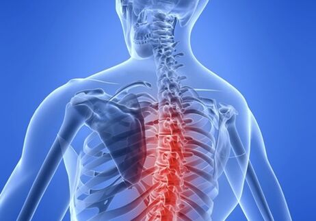
Development of thoracic osteonecrosis
With osteonecrosis, the blood supply worsens, and the transport of water, glucose and amino acids to the nuclear bone marrow, which is needed for it to synthesize water-bound carbohydrates, is disrupted. The core dries out, the gel-like structure turns into a fibrous structure, resulting in a loss of elasticity and shock absorption. The load falls on the annulus and the vertebra is injured. Microscopic particles appear on the fibrous capsule, its fibers are stretched and can no longer hold the nucleus pulposus, begin to protrude towards the spinal canal - disc protrusion. When the fibrous capsule ruptures, a herniated disc mass is formed.
Cause of disease
In people over 40–45 years of age, thoracic osteonecrosis develops as a result of the body's natural aging process. This is manifested by a delay in the regeneration of cartilage and bone tissue, a decrease in collagen production, as a result of which the elasticity and strength of the ligamentous apparatus of the spine are preserved.
At a younger age, the rapid progression of osteonecrosis of the thoracic region occurs against the background of pathologies that negatively affect the condition of the cartilage and bone tissue of the spine.
- Systemic connective tissue diseases: rheumatoid arthritis, scleroderma.
- Endocrine diseases: diabetes, hypothyroidism.
- Congenital and acquired malformations of posture: scoliosis, scoliosis.
- Prolonged exposure to static and dynamic loads.
- Hereditary predisposition to cartilage deterioration.
- Thoracic spine injury.
A sedentary lifestyle, unhealthy diet, obesity, deficiency of vitamins and trace elements in the body can cause premature destruction of discs.
Pathological level
The more the discs and vertebrae are deformed, the more obvious the clinical manifestations.
Stages of disc destruction in thoracic osteonecrosis:
I stage. The disc begins to collapse due to the pulp nucleus's inability to retain the moisture it needs to repair its tissues. The fibrous ring is covered with cracks. The patient experiences periodic chest discomfort after exertion.
Phase II. The destruction of the disc continues, the fibers of the annulus are stratified, the pulp nucleus migrates into the deep fissure that has formed in the disc surface. The height of the discs decreases, the mobility of the vertebrae increases. The muscles of the back in the affected area reflexively tense, trying to limit the mobility of the thoracic region. Moderate pain.
Phase III. If the integrity of the annulus is violated, the nucleus pulposus enters the spinal canal with the formation of a herniated disc. There is compression of the structures of the spinal cord: nerve fibers, blood vessels. The vertebral bodies were also deformed, with bone tissue growing in the form of osteoblasts. The pain becomes constant, the range of motion of the thoracic spine decreases.
Stage IV. In the late stages of thoracic osteonecrosis, signs of degeneration are observed in the ligaments, muscles and other tissues surrounding the affected spinal segment. The cartilage of the discs is replaced by scar tissue. Osteoarthritis develops in other vertebral joints. The clinical picture is varied and depends on the extent of the disc damage and the location of the herniation.
If compression of the spinal cord occurs, lens syndrome, myelopathy, and other irreversible consequences develop, leading to disability.
If the problem disc is covered by fibrous tissue and the adjacent vertebrae fuse, this can move the disease into stable remission, but with partial loss of spinal function, becoming abnormal. within the affected segment area.
Stage IV. This is the final stage of the disease. Cartilage of the intervertebral discs is replaced by connective tissue, adjacent segments of the spine participate in the pathological process. Joints grow together, becoming immobile (ankylosing spondylitis). The patient's condition is very serious: severe pain not only in the neck, but also in the arm, front of the chest, between the shoulder blades, signs of a cerebrovascular accident, impaired sensitivity. This is a life-threatening condition that can lead to a stroke.
The success of the treatment depends 90% on the experience and qualifications of the doctor.
Free consultation and diagnosis of doctors
- Chiropractic
- Skin Doctor
- Bone Trainer
- Neuroscientist
After consultation, the doctor will conduct a thorough diagnosis of the entire spine and each segment. Doctors pinpoint which nerve segments and roots are involved and causing pain symptoms. Based on the results of the consultation, detailed recommendations for treatment and, if necessary, additional diagnoses are prescribed.
Signs and symptoms of thoracic osteonecrosis
Symptoms of thoracic osteonecrosis are often confused with the clinical picture of other diseases. This is due to the fact that when the spinal roots are compressed, the functions of the organs they create inside are disturbed. The work of the gastrointestinal tract, liver, pancreas, heart is disturbed.
Chest pain is not clearly localized, can be on the arm, ribs, collarbone, shoulder blade, abdomen. In terms of the nature of the pain in osteosarcoma, they resemble those of angina, acute pancreatitis or cholecystitis.
Often, the pain between the shoulder blades is accompanied by a feeling of lack of air, which many people refer to as a heart attack.
With significant and prolonged compression of the spinal roots, a serious neuropathy develops with motor and sensory disturbances. Specifically, the location of the disorder depends on which thoracic vertebra is near which nerve root.
The area of pain and sensitivity changes in the form of numbness extending from the neck, shoulder blades, ribs, breastbone to the abdomen.
Principles of disease diagnosis
Diagnosis of osteonecrosis involves the following steps:
- Prehistoric Collection.
- Clinical examination with assessment of neurological status.
- function tests.
- Measurement methods: X-ray, magnetic resonance and computed tomography.
An important stage of the examination is the differential diagnosis. Symptoms of thoracic osteonecrosis are often "disguised" as heart, stomach, and lung diseases, so additional research methods are indicated for an accurate diagnosis.
The treatment
Most patients with signs of osteonecrosis of the thoracic spine require conservative treatment. Surgical treatment is performed only in particularly severe cases, when the spinal canal is significantly narrowed due to a hernia and the spinal cord is severely compressed.
In modern clinics for the treatment of osteonecrosis, the author's methods are used non-surgically, which not only allow to eliminate pain in the acute period, but also stabilize the condition of the spine, preventprevent the development of complications. For each patient, depending on the severity of the pathology, a treatment strategy is selected.
Steroid mastoid tumor: symptoms and treatment of spina bifida in a modern clinic
The goals of drug therapy for osteonecrosis:
- Mass pain syndrome.
- Reduce inflammation.
- Normalizes metabolism.
- Improves blood supply.
- Reduce muscle spasms.
Drugs used: anesthetics, anti-inflammatory drugs, steroid hormones, muscle relaxants, B vitamins.
Modern medical centers have improved the classic manual therapy methods, adding electrophoresis and photodynamic laser therapy to improve treatment efficiency.
Therapy includes:
- Soft manual techniques work at the physiological level and allow you to successfully remove the pinched nerve roots in the spine.
- Multicomponent electrophoresis is a medical procedure in which drugs are directly injected into the lesion.
- Laser therapy. Under the impact of laser radiation, topical drugs in the damaged spine will penetrate 10-15 cm deep and have analgesic and anti-inflammatory effects at the cellular level.
Vertebral blockade is a method of putting anesthetic into the damaged nerve root area, helping to quickly eliminate pain, reduce swelling, inflammation, and replenish blood.
Shockwave therapy, in which sound vibrations at a certain frequency cause an effect similar to an electric massage. The therapeutic effect of the procedure lies in its analgesic effect and enhanced tissue regeneration.
Physical therapy exercises, strengthening the back muscles, contribute to the formation of a naturally strong corset that helps maintain the spine in the correct anatomical position.
Years of experience in treating thoracic spine tumors in a professional clinic show that symptoms that complicate patients' lives, with the right and comprehensive treatment, disappear, preventing progression. further development of the pathological process.